Tissue Doppler Imaging is an advanced ultrasound technique designed to measure the velocity of moving tissues within the heart. Unlike conventional Doppler imaging, which primarily focuses on blood flow, TDI zeroes in on the motion of the heart's muscle tissues. This provides an in-depth analysis of cardiac function, contributing to a more comprehensive understanding of the heart's performance.
Mechanism of
Tissue Doppler Imaging
During
a TDI examination, ultrasound waves are directed toward the heart, and the
returning signals are meticulously analyzed to create detailed images of tissue
movement. By gauging the speed and direction of these movements, we can
identify abnormalities in the contraction and relaxation of the heart muscle.
TDI
is instrumental in providing crucial insights into:
·
Myocardial Function: A detailed
evaluation of the heart muscle's function, identifying areas that may exhibit
weakness or compromise.
·
Valvular Function: Assessment of the
movement and function of heart valves, aiding in the detection of regurgitation
or stenosis.
·
Dyssynchrony: Identification of asynchrony in the
contraction of different segments of the heart muscle, which can be indicative
of certain cardiac conditions.
The Advantages
of Tissue Doppler Imaging
·
Early Detection: TDI facilitates the early detection of
subtle changes in cardiac function, enabling prompt intervention and the
development of personalized treatment plans.
·
Increased Sensitivity: TDI offers greater
sensitivity in detecting abnormalities compared to traditional imaging methods,
providing a more detailed assessment of the heart.
·
Quantitative Analysis: The technique
provides quantitative data on myocardial velocities, allowing for precise and
objective measurements in the evaluation of cardiac function.
Applications
of Tissue Doppler Imaging
Tissue
Doppler Imaging finds applications in various clinical scenarios, including:
·
Congestive Heart Failure Management
·
Coronary Artery Disease Assessment
·
Valvular Heart Disease Evaluation
·
Cardiomyopathy Diagnosis and Monitoring
Detailed
Information on Tissue Doppler Imaging
1.
Practical
Aspects of the TDI Procedure:
·
Preparation: Before the
procedure, patients may be asked to refrain from eating or drinking for a
specified period. Comfortable clothing is recommended.
·
Procedure: During the TDI
examination, a transducer is placed on the chest, and ultrasound waves are
directed toward the heart. The process is painless and typically takes about 30
to 60 minutes.
·
Post-Procedure: There is
usually no downtime, and patients can resume normal activities immediately
after the examination.
2.
Interpreting
TDI Results:
·
Velocity Patterns: TDI
produces velocity patterns that are analyzed to assess the motion of different
regions of the heart.
·
Abnormal Findings: The
examination helps identify abnormalities such as reduced myocardial velocities,
dyssynchrony, and impaired valvular function.
·
Clinical Correlation: TDI
results are correlated with clinical data and other diagnostic findings to
provide a comprehensive understanding of the patient's cardiac health.
3.
Patient
Education and Engagement:
·
Education: Our team believes in
empowering patients with knowledge. We provide detailed explanations of TDI
procedures and results to ensure patients are well-informed about their cardiac
health.
·
Engagement: Patients are
encouraged to actively participate in their healthcare journey, ask questions,
and communicate any concerns they may have.
4.
Follow-Up
Care and Monitoring:
·
Customized Care Plans: Based on
TDI findings, our medical team develops customized care plans tailored to each
patient's unique needs.
·
Regular Follow-Up: Periodic
follow-up appointments and monitoring are scheduled to track progress, assess
treatment effectiveness, and make adjustments as necessary.
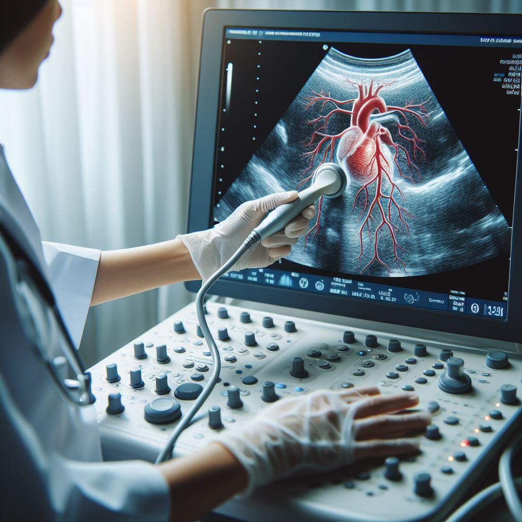
Doppler Imaging Procedure
The Tissue Doppler Imaging (TDI) procedure is a non-invasive and painless
diagnostic test that provides valuable insights into the functioning of your
heart muscle. Here's what you can expect during the TDI examination at
[Doctor's Name] Clinic:
1.
Preparation: You may be asked to change into a hospital gown to allow easy access to
the chest area. In some cases, small adhesive patches called electrodes may be
placed on your chest to monitor your heart's electrical activity.
2.
Positioning: You will lie comfortably on an examination table, and a trained
sonographer will apply a special gel to the skin over your chest. This gel
helps to transmit sound waves and improve the quality of the images.
3.
Transducer Placement: The transducer, a small handheld device, will be moved gently over the
chest. The transducer emits sound waves, and the returning signals are
converted into detailed images of the heart's tissue movements.
4.
Image Acquisition: The sonographer will focus on specific areas of the heart, capturing
images that reflect the velocity and direction of tissue movements. The procedure
may take approximately 30 to 60 minutes, depending on the complexity of the
evaluation.
5.
Analysis: The collected
images will be carefully analyzed by our skilled healthcare team, including
[Doctor's Name], to assess myocardial function, valvular movement, and any
signs of dyssynchrony.
Risks and Considerations
Tissue Doppler Imaging is generally considered a safe diagnostic
procedure with minimal risks. However, it's essential to be aware of the
following considerations:
1.
No Radiation Exposure: TDI uses ultrasound technology, eliminating the risk associated with
radiation exposure commonly found in other imaging techniques.
2.
Potential Discomfort: The procedure is generally well-tolerated, but you may experience mild
discomfort from the pressure of the transducer on your chest or the coolness of
the gel.
3.
Allergic Reactions: The gel used during the procedure is typically hypoallergenic. However,
if you have a known allergy to ultrasound gel, please inform our healthcare
team before the examination.
4.
Limited Diagnostic Scope: While TDI provides valuable information about cardiac tissue movements,
it may not be suitable for certain conditions. Your healthcare provider will
determine the most appropriate diagnostic approach based on your individual
needs.
It's important to communicate openly
with our healthcare team about any pre-existing medical conditions, allergies,
or concerns you may have before undergoing Tissue Doppler Imaging. Your comfort
and well-being are our top priorities, and we are here to address any questions
or uncertainties you may have.
All Services
-
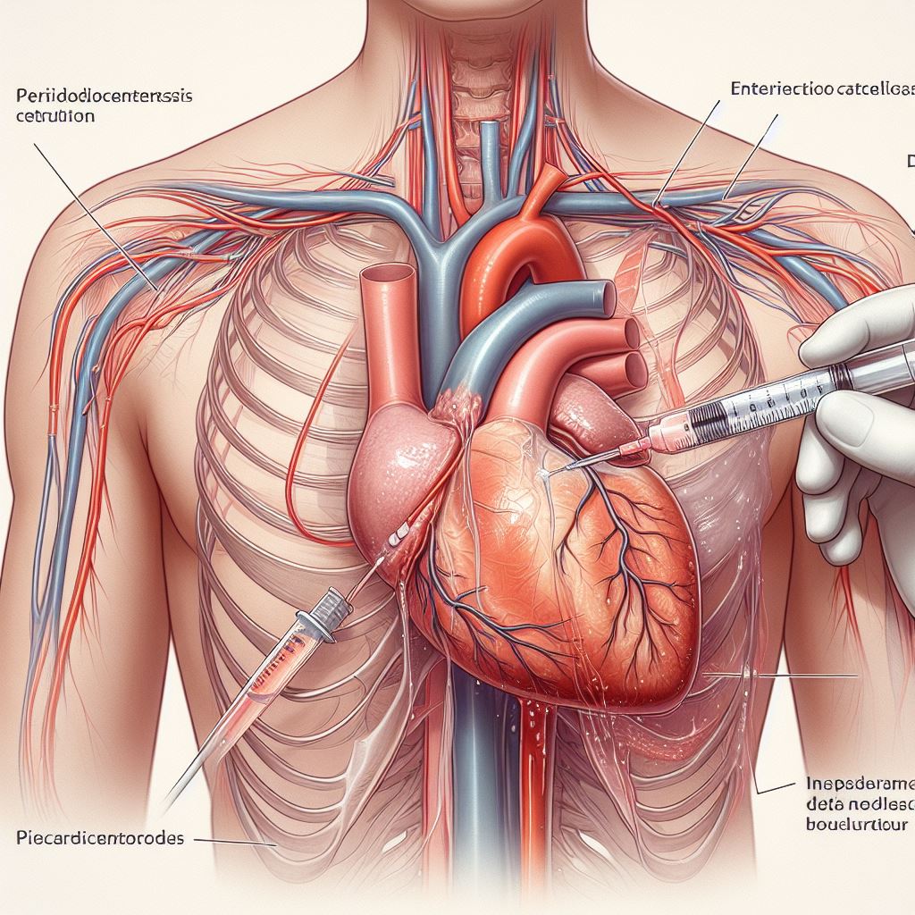 Pericardiocentesis
-
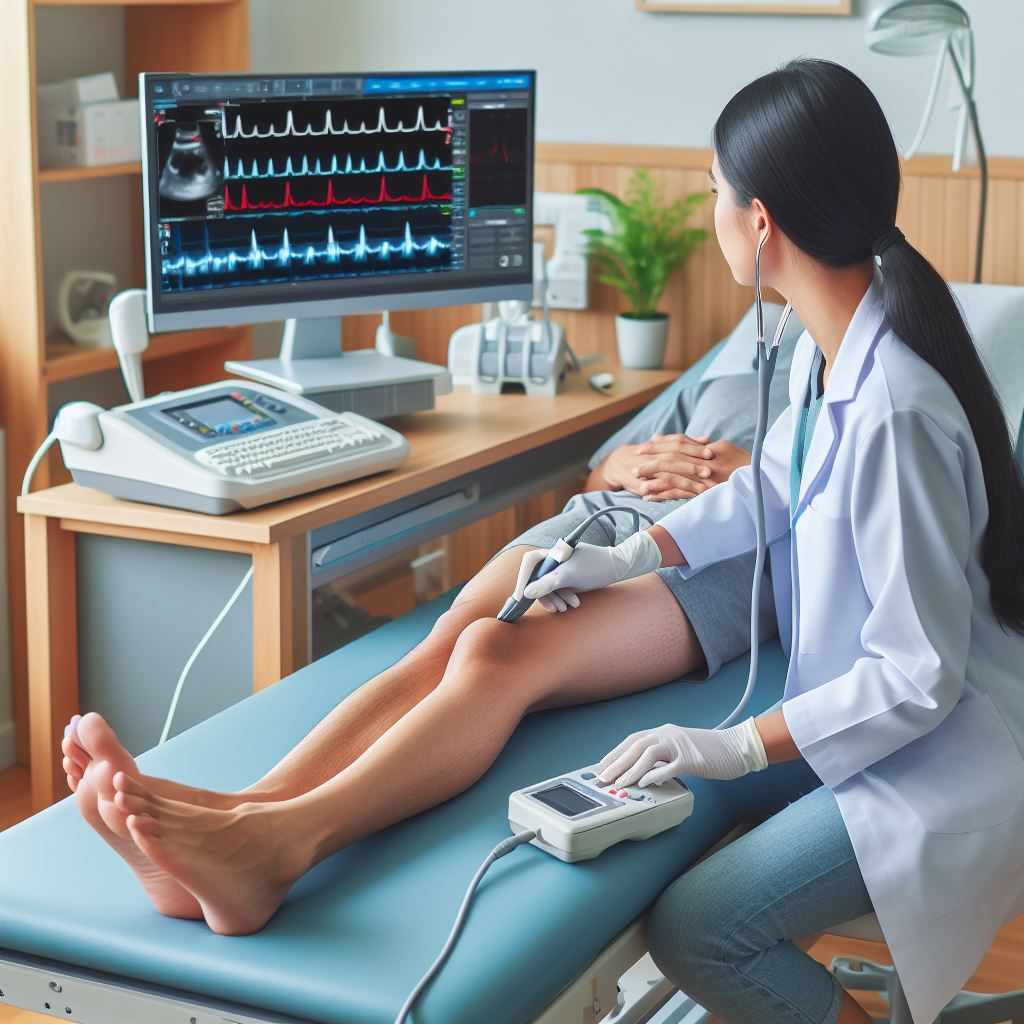
 Echocardiography
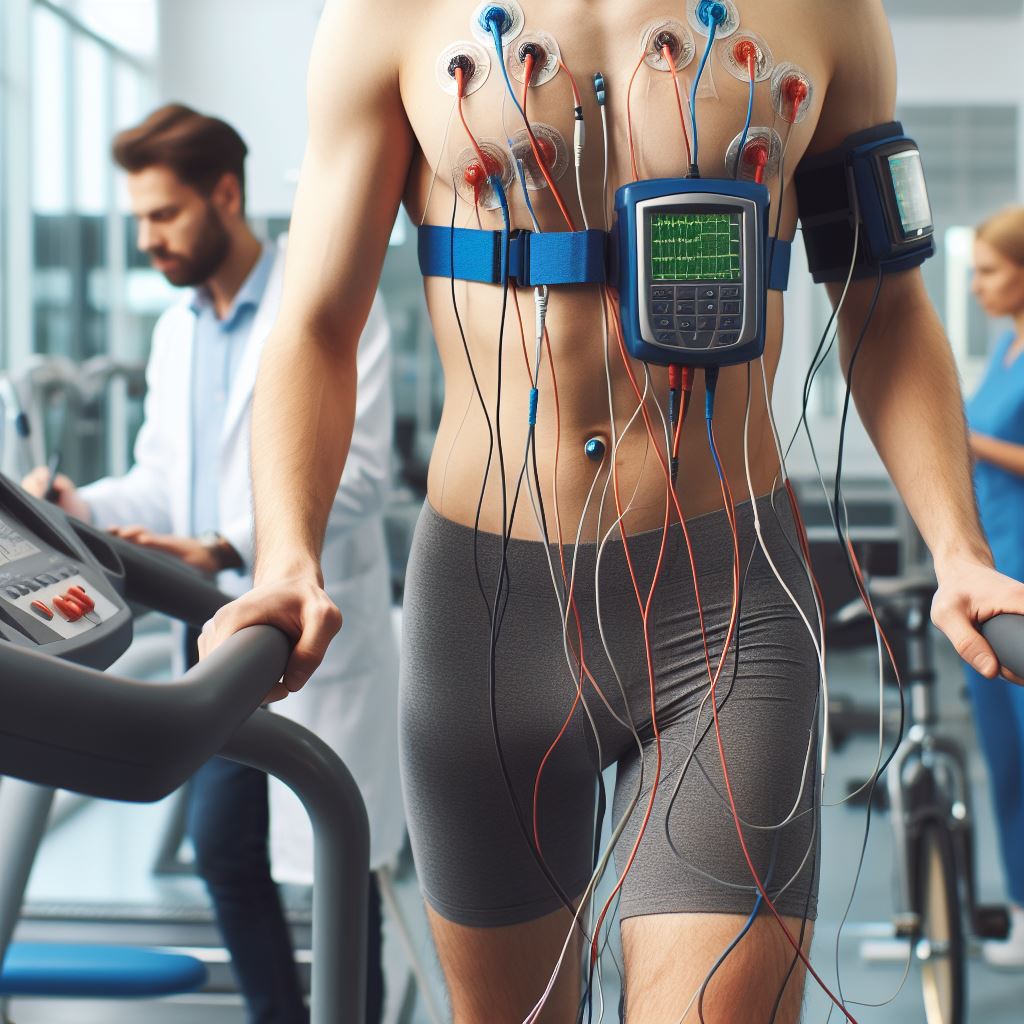
-
 Treadmill Stress Testing
-
 Holter Monitoring
-
 2D Echocardiography & Doppler Studies
-
Tissue Doppler Imaging
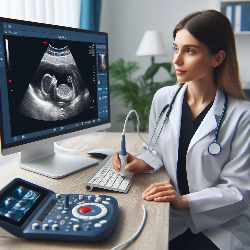
-
 Fetal Echocardiography
-
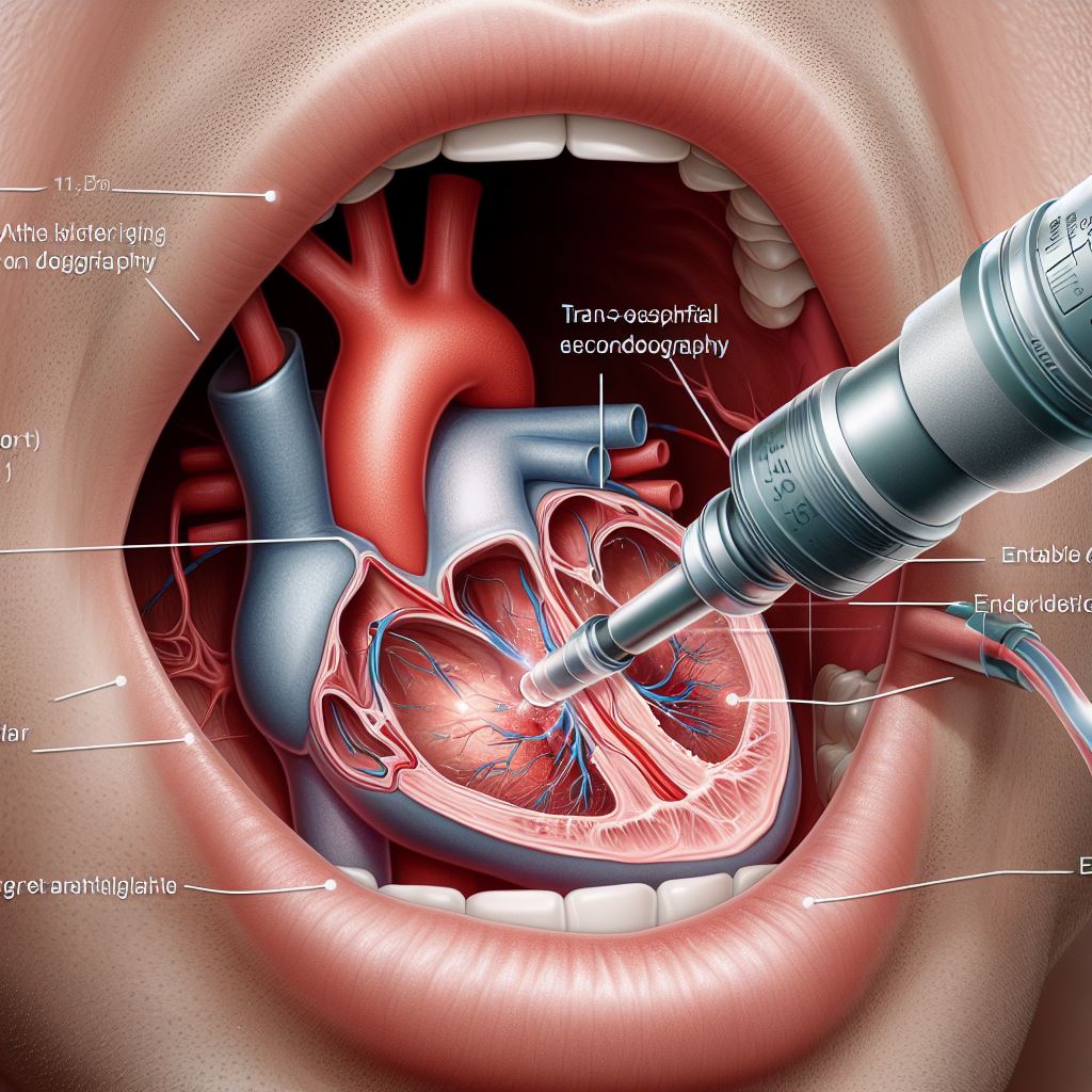 Trans-Oesophageal Echocardiography (TOE)
-
 Peripheral and Carotid Doppler Studies
-
 Stress Echocardiography
-
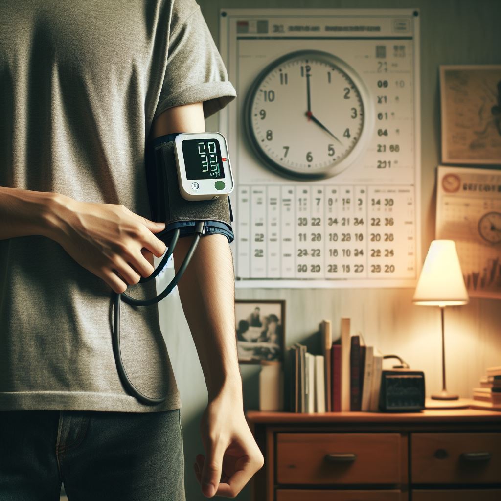 Ambulatory Blood Pressure Monitoring
-
 CT Imaging
-
 Sudden Cardiac Arrest