
Fetal Echocardiography is a specialized ultrasound that focuses on the
development and function of the fetal heart. This non-invasive imaging
technique allows doctors to examine the structure and function of the baby's
heart in the womb, offering crucial insights into potential cardiac
abnormalities. The heart is a complex organ, and any issues detected early in
pregnancy can significantly impact treatment and management strategies.
When is Fetal Echocardiography Used?
This procedure is typically recommended in the following situations:
1.
Maternal Risk Factors: If the mother has a history of congenital heart disease, exposure to
medications or substances that may impact fetal heart development, or a
previous child with heart abnormalities.
2.
Abnormalities Detected in Routine
Ultrasound: If a routine ultrasound identifies potential
issues with the baby's heart structure or function.
3.
Family History of Heart Conditions: If there is a family history of congenital heart defects or genetic
syndromes associated with heart problems.
4.
Maternal Diabetes: Due to the increased risk of cardiac abnormalities in babies born to
mothers with diabetes.
Early detection of heart
abnormalities allows for timely intervention and planning, improving the
chances of a positive outcome for both the mother and the baby.
Do I Need to Prepare for the Procedure?
Fortunately, no special preparation is required for fetal
echocardiography. It is a safe and non-invasive procedure that does not involve
any radiation. Mothers are encouraged to eat and drink normally before the
exam. However, it's essential to communicate any existing health conditions or
concerns with your healthcare provider before the procedure.
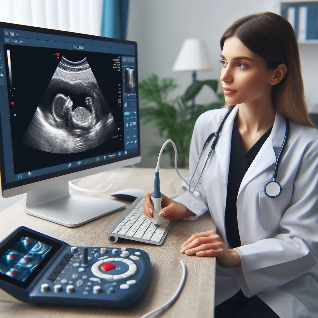
What Happens During the Exam?
The procedure involves placing a gel on the mother's abdomen and using a
transducer to emit sound waves that create detailed images of the fetal heart.
The exam is painless and typically takes about 30 to 60 minutes, depending on
the complexity of the evaluation. The transducer is moved around the abdomen to
capture different angles and views of the baby's heart. High-frequency sound
waves bounce off the structures in the heart, and a computer translates these
echoes into images.
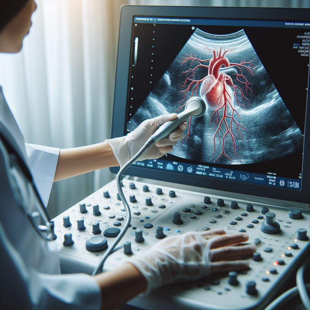
Advanced imaging technology, such as
3D and 4D ultrasound, may also be used to provide even more detailed images of
the fetal heart. This allows for a comprehensive assessment of the heart's
structure, function, and blood flow.
Are There Any Risks Associated with This Exam?
Fetal echocardiography is considered safe, with no known risks to the
mother or the baby. It is an essential tool for early detection and management
of potential heart issues. The benefits of obtaining critical information about
the baby's heart health far outweigh any minimal risks associated with the
procedure.
The procedure is conducted by trained
and experienced healthcare professionals who prioritize the safety and
well-being of both the mother and the baby. It is crucial to follow the
guidance of your healthcare provider and attend scheduled prenatal appointments
for a thorough assessment of your baby's development.
What Do the Results Mean?
The results of the fetal echocardiography are carefully analyzed by our
skilled team of cardiologists. Normal results provide reassurance, indicating
that the baby's heart is developing as expected. In cases where abnormalities
are detected, a detailed discussion with the parents is initiated.
Understanding the nature of the abnormalities allows for the development of a
comprehensive care plan for the baby.
The information obtained from the
fetal echocardiography can help healthcare providers plan for potential
interventions, surgeries, or specialized care that may be needed after the baby
is born. Early diagnosis enables the medical team to collaborate with parents
and provide the necessary support and resources for the best possible outcome.
Why is This Test Important?
Fetal echocardiography is crucial for several reasons:
1.
Early Detection and Intervention: Early identification of heart abnormalities allows for timely medical
intervention and planning. This can significantly improve the chances of a
positive outcome for the baby.
2.
Tailored Care Plans: Knowledge of potential cardiac issues enables healthcare providers to
create individualized care plans that address the specific needs of the baby
and the family.
3.
Emotional Support: The emotional well-being of expectant parents is a vital aspect of
prenatal care. Fetal echocardiography provides reassurance for parents when the
results are normal and allows for early preparation and support when
abnormalities are detected.
4.
Coordination of Care: The information obtained from fetal echocardiography facilitates
coordination between different medical specialists involved in the care of the
baby, ensuring a seamless and collaborative approach to treatment.
Benefits
1.
Early Detection of Heart Abnormalities: Fetal echocardiography allows for the early identification of
structural or functional issues with the baby's heart.
2.
Tailored Care Plans: Information from the procedure enables healthcare providers to create
personalized care plans that address the specific needs of the baby.
3.
Emotional Reassurance: Normal results provide emotional reassurance for expectant parents,
while early detection of abnormalities allows for proactive planning and
support.
All Services
-
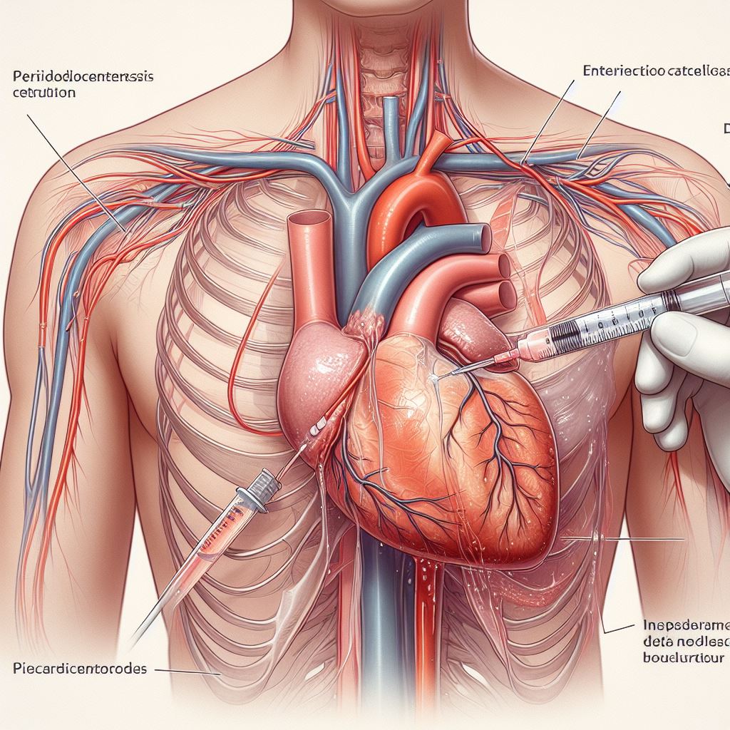 Pericardiocentesis
-
 Echocardiography
-
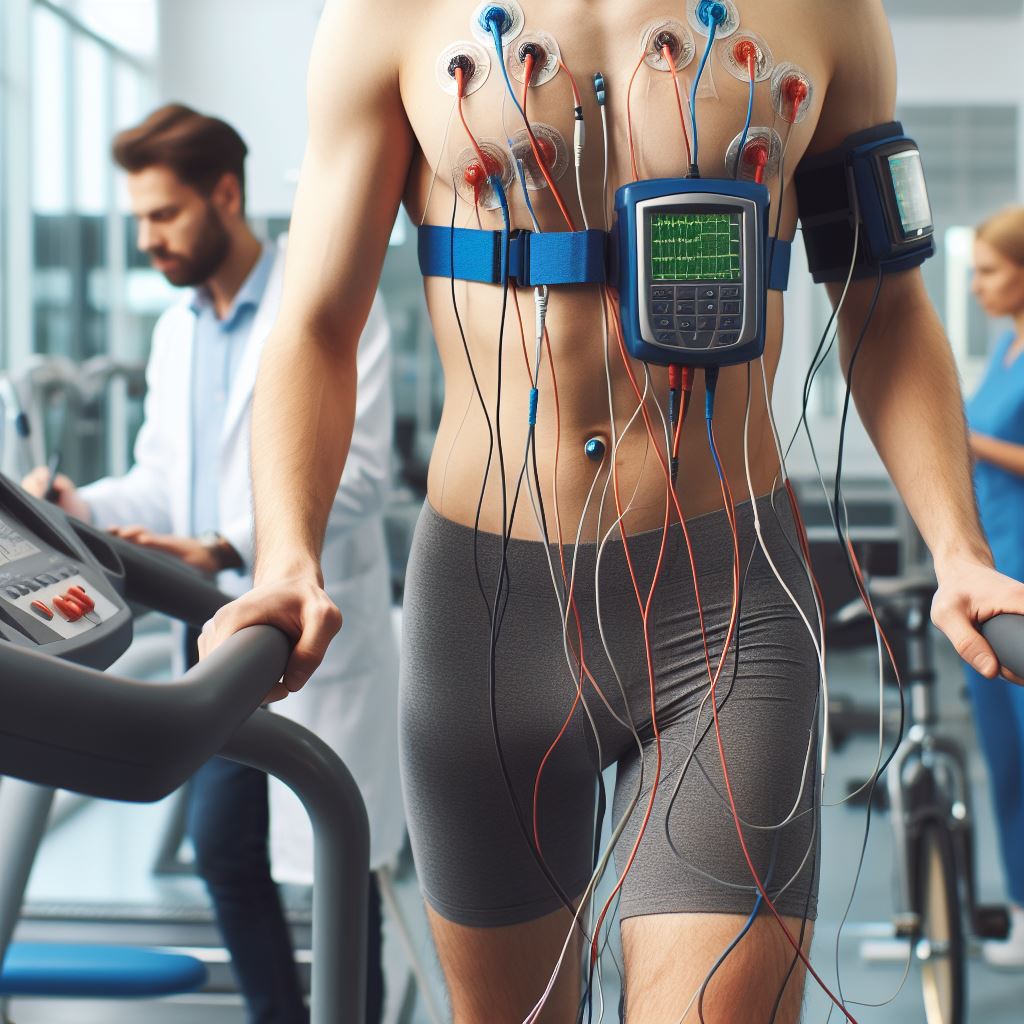
 Treadmill Stress Testing
-
 Holter Monitoring
-
 2D Echocardiography & Doppler Studies
-
 Tissue Doppler Imaging
-
Fetal Echocardiography
-
 Trans-Oesophageal Echocardiography (TOE)
-
 Peripheral and Carotid Doppler Studies
-
 Stress Echocardiography
-
 Ambulatory Blood Pressure Monitoring
-
 CT Imaging
-
 Sudden Cardiac Arrest