
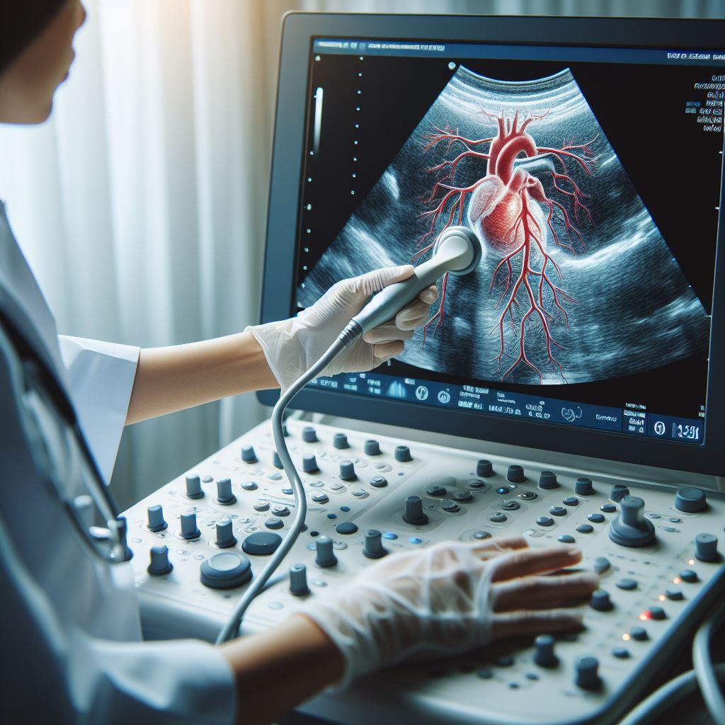
Echocardiography
Overview of Echocardiography:
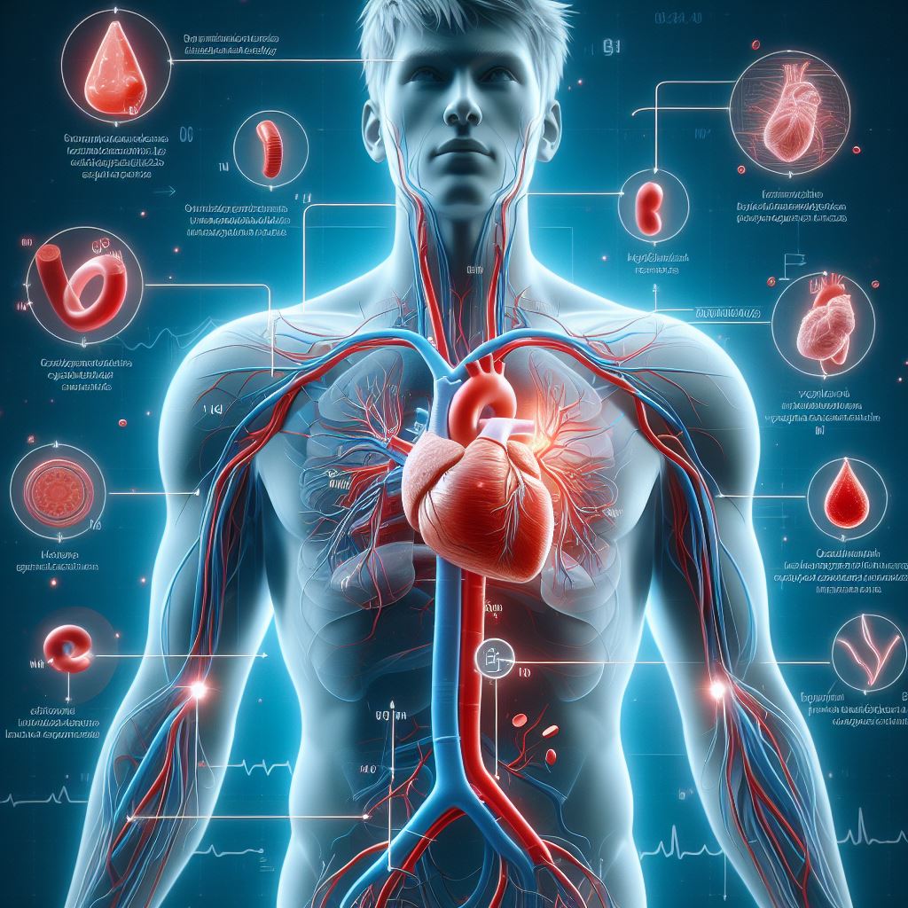
Echocardiography stands as a cornerstone in the field of cardiac
imaging, providing detailed insights into the structure and function of the
heart. This non-invasive procedure employs ultrasound waves to create real-time
images, enabling healthcare providers to assess cardiac health comprehensively.
Types of Echocardiograms:
1. Transthoracic Echocardiogram (TTE):
·
TTE is the most prevalent type,
involving the placement of a transducer on the chest. It captures images of the
heart by sending and receiving ultrasound waves through the chest wall.
2. Transesophageal Echocardiogram (TEE):
·
TEE offers a more detailed view by
inserting a transducer into the esophagus, providing clearer images of the
heart's posterior structures.
3. Doppler Echocardiogram:
·
Doppler technology, an extension of
echocardiography, evaluates blood flow within the heart and blood vessels. It
provides crucial information about circulation and helps identify
abnormalities.
Echocardiogram Methods:
Understanding the basic procedure is integral for patients:
· Gel Application: A gel is applied to the chest or transducer to enhance the transmission
of sound waves.
· Transducer Placement: The transducer is moved across the chest or inserted into the esophagus
to capture images from different angles.
· Real-time Imaging: Results are displayed in real-time on a monitor, allowing immediate
assessment and analysis.
Why It's Performed:
Echocardiography serves various diagnostic purposes:
· Assessment of Heart Function: It evaluates the heart's pumping capacity and overall function, aiding
in the diagnosis of heart failure or other functional abnormalities.
· Identification of Structural Abnormalities: Echocardiography detects structural issues such as
valve disorders, congenital heart defects, and cardiac masses.
· Monitoring Heart Conditions: The procedure is instrumental in tracking the progression or
improvement of known heart conditions, ensuring timely intervention when
necessary.
How to Prepare:
Patients can take specific steps to prepare for an echocardiogram:
· Clothing Choice: Wear comfortable clothing, as you may be asked to remove your upper
garments.
· Fasting Instructions: Depending on the type of echocardiogram, fasting for a few hours before
the procedure might be required.
· Adherence to Instructions: Follow any specific instructions provided by your healthcare provider,
ensuring optimal conditions for the procedure.
Risks:
While echocardiography is generally considered safe, there are minimal
risks associated with the procedure:
· Allergic Reactions: In rare instances, patients may experience allergic reactions to the
ultrasound gel used during the procedure.
· Discomfort with TEE: Some discomfort may be encountered during a transesophageal
echocardiogram, as the transducer is inserted into the esophagus.
Results:
Interpreting echocardiogram results is a crucial aspect of cardiac care.
Your healthcare provider will analyze the findings and discuss them with you,
offering valuable insights into your heart health.
The results of an echocardiogram may
encompass:
· Structural Information: The images provide detailed information about the structure of the
heart, including the chambers, valves, and surrounding tissues.
· Functional Assessment: Echocardiography assesses how well the heart is pumping blood,
providing vital information about its overall function.
· Blood Flow Dynamics: Doppler echocardiography evaluates blood flow patterns within the heart
and blood vessels, aiding in the detection of abnormalities.
Understanding Echocardiography Results:
1. Normal Findings:
·
A normal echocardiogram indicates
that the heart is functioning within the expected parameters, with no
significant structural abnormalities.
2. Abnormal Findings:
·
Abnormalities may include issues such
as valve disorders, weakened heart muscles, congenital anomalies, or signs of
heart disease.
3. Follow-up Recommendations:
·
Depending on the findings, your
healthcare provider may recommend further tests, lifestyle modifications, or
specific treatments to address identified issues.
Importance of Communication:
Open communication between patients and healthcare providers is paramount:
· Questions and Concerns: Don't hesitate to ask questions or express concerns about the procedure
or its results.
· Follow-up Discussions: Schedule follow-up appointments to discuss results and develop an
appropriate plan of action based on the findings.
· Collaborative Decision-making: Work collaboratively with your healthcare team to make informed
decisions about your cardiac health.
All Services
-
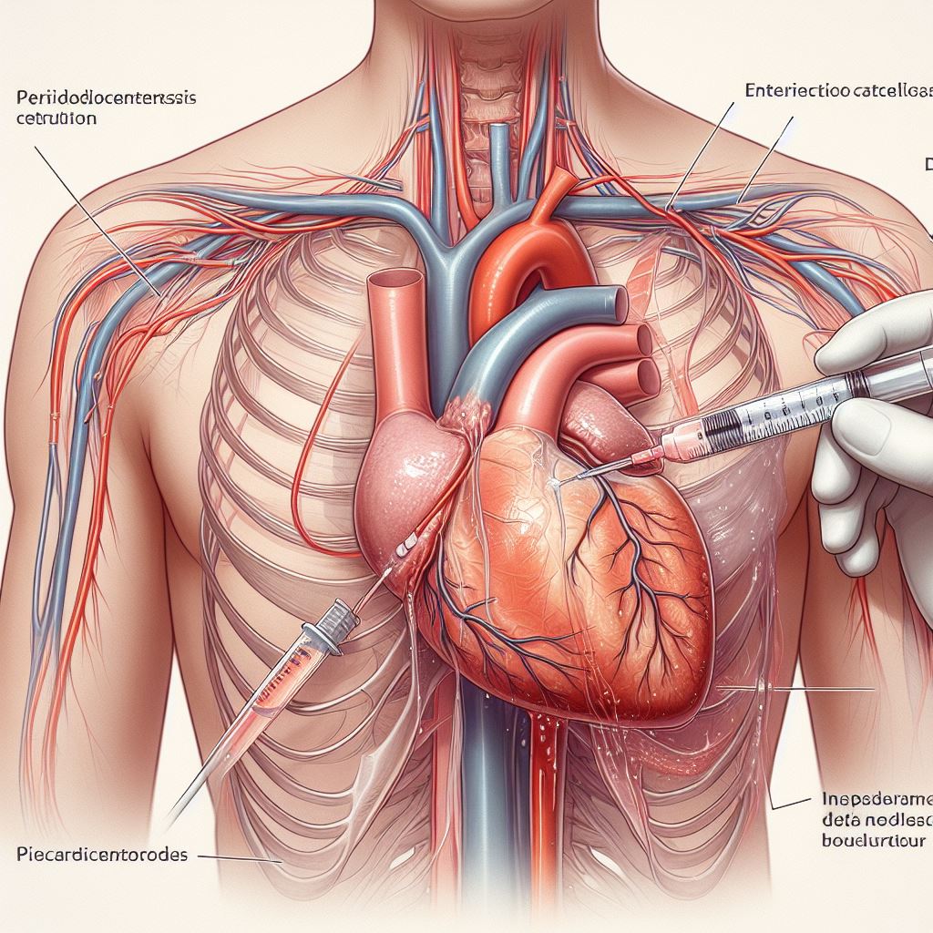 Pericardiocentesis
-
Echocardiography
-
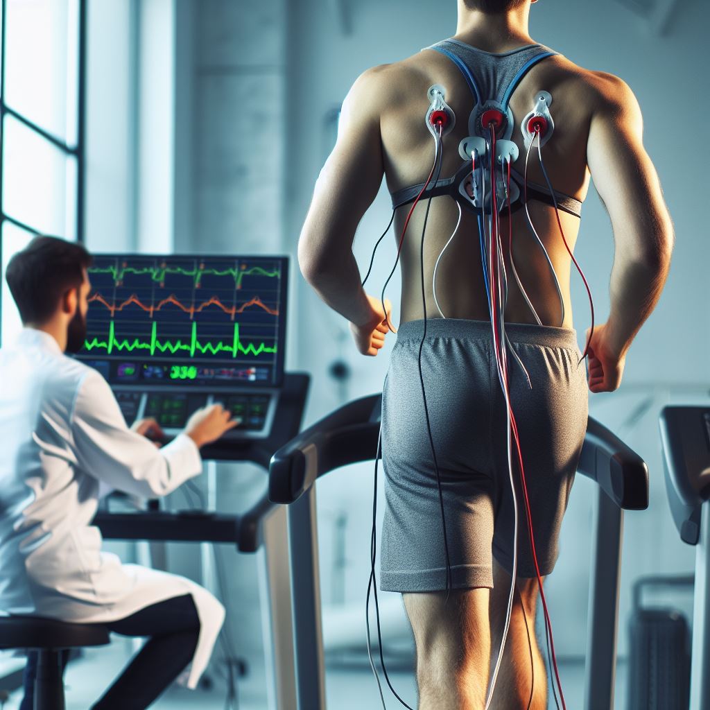 Treadmill Stress Testing
-
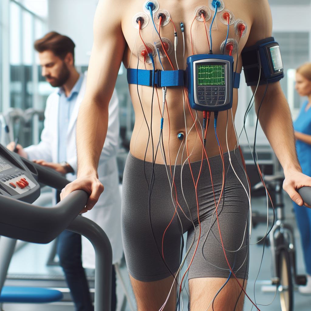 Holter Monitoring
-
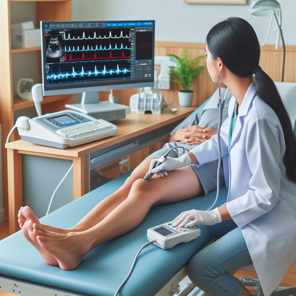
 2D Echocardiography & Doppler Studies
-
 Tissue Doppler Imaging
-
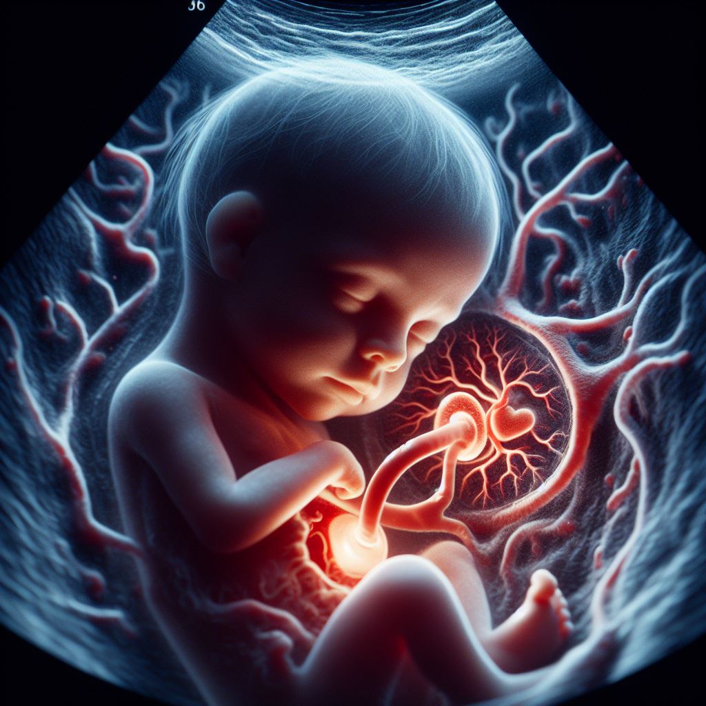 Fetal Echocardiography
-
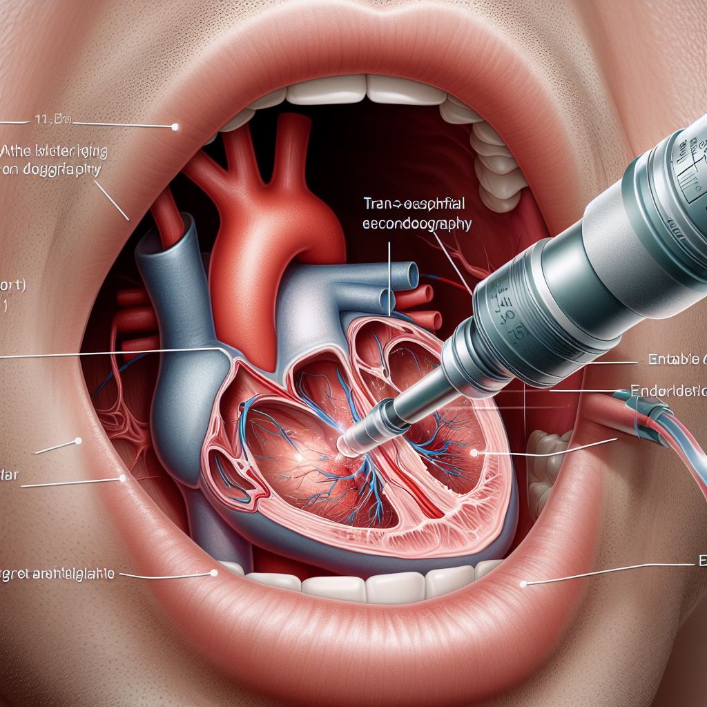 Trans-Oesophageal Echocardiography (TOE)
-
 Peripheral and Carotid Doppler Studies
-
 Stress Echocardiography
-
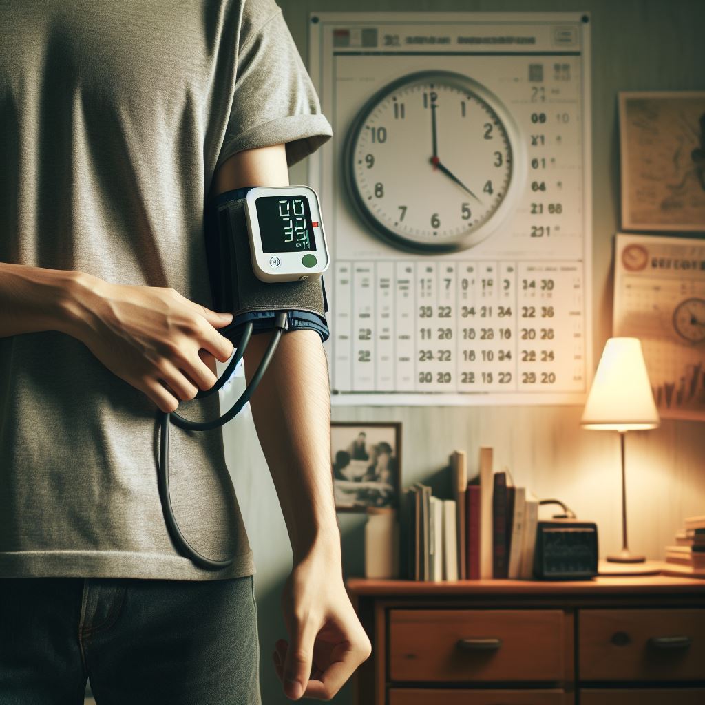 Ambulatory Blood Pressure Monitoring
-
 CT Imaging
-
 Sudden Cardiac Arrest