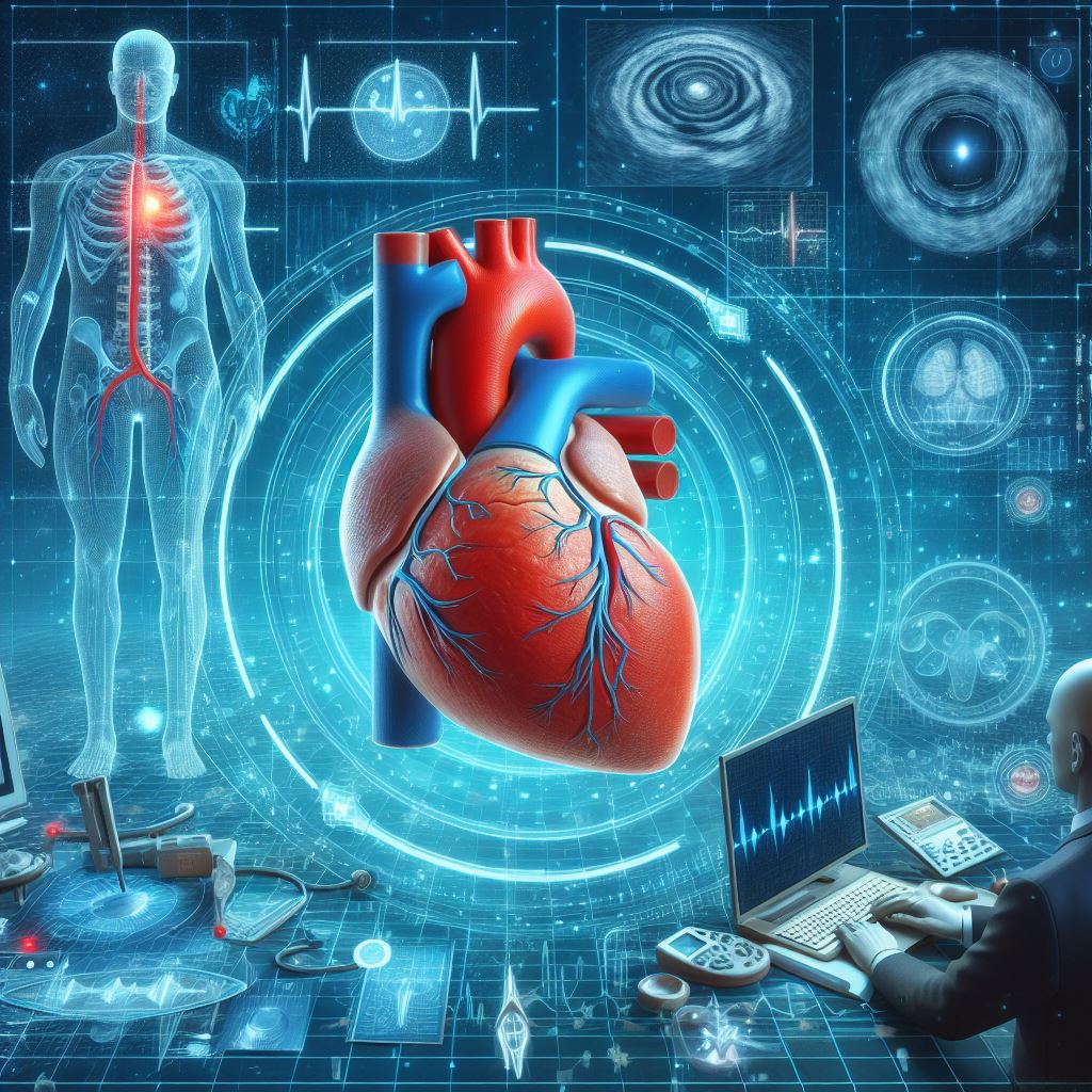
In the ever-evolving landscape of
cardiovascular health, our cardiology center embraces advanced diagnostic tools
to deliver superior patient care. Among these tools, 2D Echocardiography stands
out as a pivotal non-invasive imaging technique that provides unparalleled
insights into the structure and function of the heart. This sophisticated
technology employs ultrasound waves to generate two-dimensional images,
enabling our expert cardiologists to meticulously examine the heart's chambers,
valves, and overall performance.
Why Should You Undergo a 2D Echo?
1. Early Detection and Prevention:
· Heart Valve Diseases: 2D Echocardiography plays a crucial role in the early identification of
heart valve diseases, allowing for timely interventions that can significantly
impact outcomes.
· Congenital Heart Defects: By capturing detailed images, this diagnostic tool aids in the
detection of congenital heart defects in both children and adults, facilitating
early management and personalized treatment plans.
· Structural Abnormalities: Assessing the heart's size and shape is vital for identifying structural
abnormalities that may contribute to various cardiovascular conditions. Early
detection enables proactive measures to prevent the progression of these
issues.
2. Monitoring Heart Conditions:
· Progression Monitoring: For individuals with known heart conditions, regular 2D
Echocardiography is indispensable for monitoring disease progression. This
ongoing assessment allows our cardiologists to make informed decisions about
adjusting treatment plans and ensuring optimal patient care.
· Treatment Evaluation: By providing real-time images, 2D Echocardiography aids in evaluating
the effectiveness of ongoing treatments. This iterative process allows for
adjustments in treatment strategies, ensuring patients receive the most beneficial
and personalized care.
Procedure: What to Expect During a 2D Echo
Preparation: Before the procedure, minimal preparation is typically required. You
may be asked to change into a gown, and small adhesive patches (electrodes)
will be placed on your chest to record your heart's electrical activity.
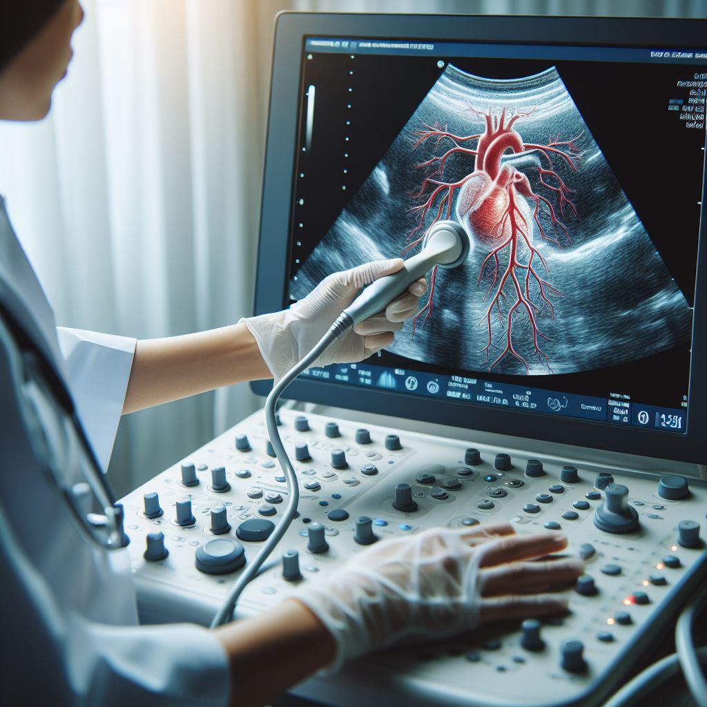
During the Test: The 2D Echocardiography procedure is performed in a controlled
environment. You will lie on an examination table, and a skilled sonographer
will apply a special gel to your chest. The transducer, a small hand-held
device, will be moved across various angles of your chest to capture detailed
images of your heart. This painless procedure typically lasts between 30 to 60
minutes, providing a comprehensive examination of your cardiac structure and
function.
Risks and Benefits
Benefits:
· Non-Invasive Nature: 2D Echocardiography is non-invasive, eliminating the need for surgical
procedures or exposure to ionizing radiation. This characteristic ensures
patient safety and comfort during the diagnostic process.
· Accurate Diagnosis: The real-time imaging capability of 2D Echocardiography allows for
accurate and detailed diagnoses of a wide range of cardiac conditions. This
precision is instrumental in tailoring treatment plans to individual patient
needs.
· Guidance for Treatment Decisions: The insights gained from 2D Echocardiography guide healthcare
professionals in making informed decisions about treatment strategies, ensuring
that interventions are targeted and effective.
· Monitoring Interventions: Following interventions or treatments, 2D Echocardiography is
invaluable for monitoring the impact and success of these measures. This
proactive approach allows for timely adjustments to optimize patient outcomes.
Risks: While 2D
Echocardiography is considered a safe diagnostic tool with minimal risks, it is
essential to acknowledge potential considerations:
· Allergic Reactions: In rare instances, individuals may exhibit allergic reactions to the
gel used during the procedure. However, such reactions are exceptionally
uncommon.
· Discomfort from Gel: Some patients may experience mild discomfort due to the gel's
temperature, but this is generally temporary and does not pose significant
challenges.
Recovery and Follow-Up
Following a 2D Echocardiography,
there is typically no downtime, and you can resume your regular activities
immediately. The images captured during the procedure will be meticulously
reviewed by our experienced cardiologists, and the findings will be discussed
with you in detail.
Post-Procedure Guidance:
· Consultation: Your cardiologist will schedule a follow-up consultation to discuss the
results of the 2D Echocardiography. This comprehensive discussion ensures that
you have a clear understanding of your cardiac health status.
· Further Testing or Treatments: If additional tests or treatments are deemed necessary based on the
findings, a personalized plan will be developed. This may include additional
imaging studies, medication adjustments, or if needed, invasive procedures.
At our cardiology
center, patient education and comfort are paramount. We recognize that
understanding your cardiac health is essential for making informed decisions
about your well-being. Our dedicated team of cardiologists and healthcare
professionals is available to address any questions or concerns you may have
about 2D Echocardiography or any other cardiac procedures. Your heart health is
our priority, and we are committed to providing the highest standard of care to
ensure your cardiovascular well-being.
All Services
-
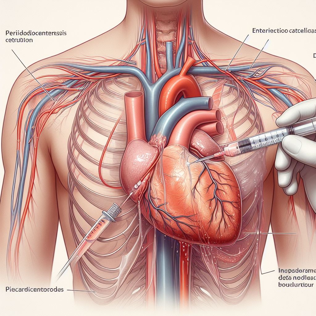 Pericardiocentesis
-
 Echocardiography
-
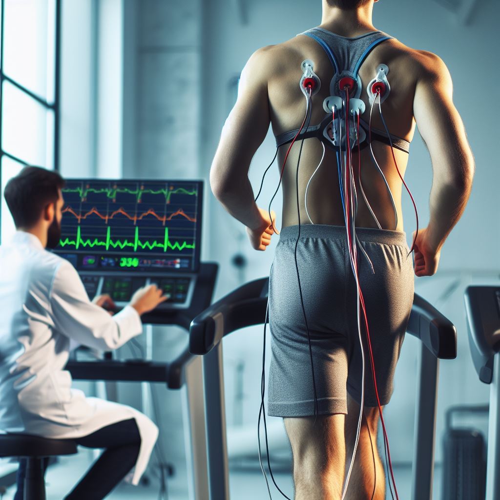 Treadmill Stress Testing
-
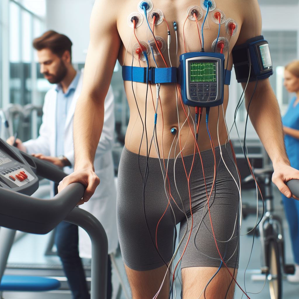 Holter Monitoring
-
2D Echocardiography & Doppler Studies
-
 Tissue Doppler Imaging
-
 Fetal Echocardiography
-
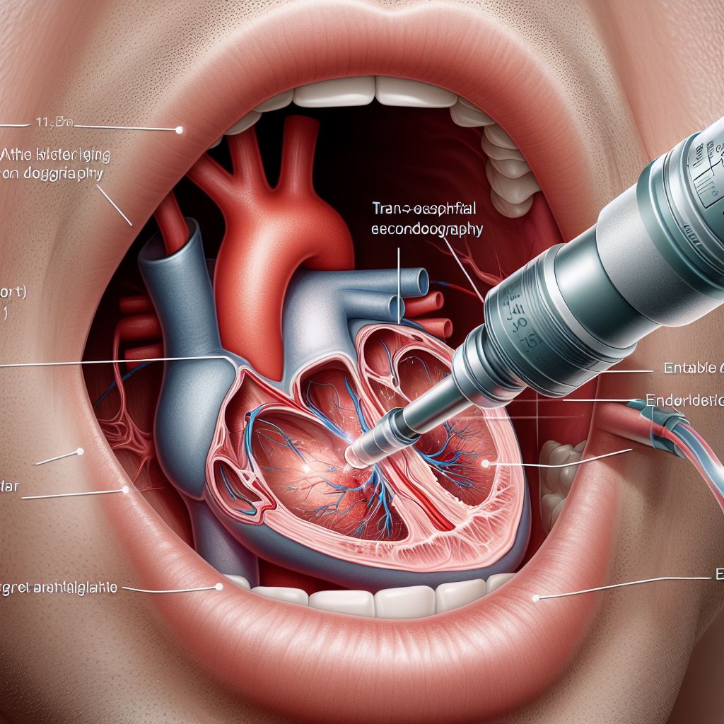 Trans-Oesophageal Echocardiography (TOE)
-
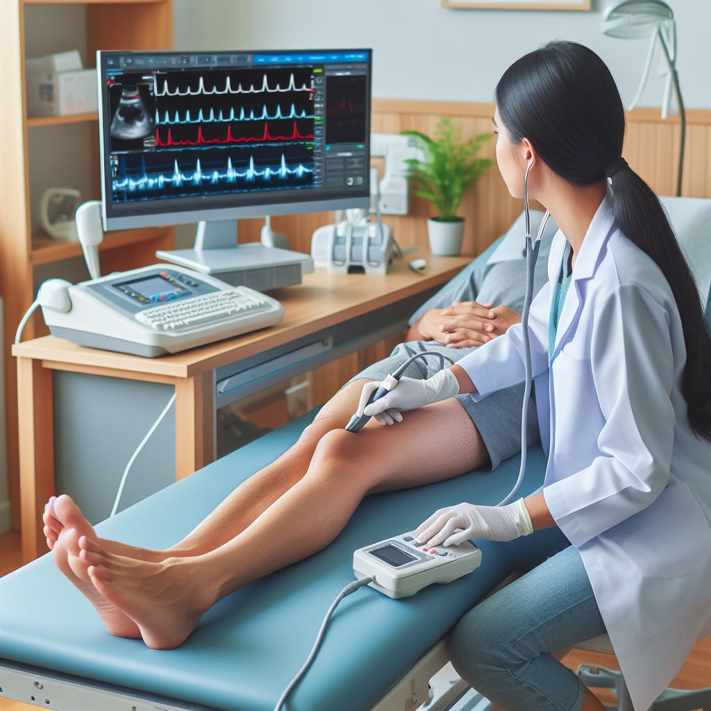 Peripheral and Carotid Doppler Studies
-
 Stress Echocardiography
-
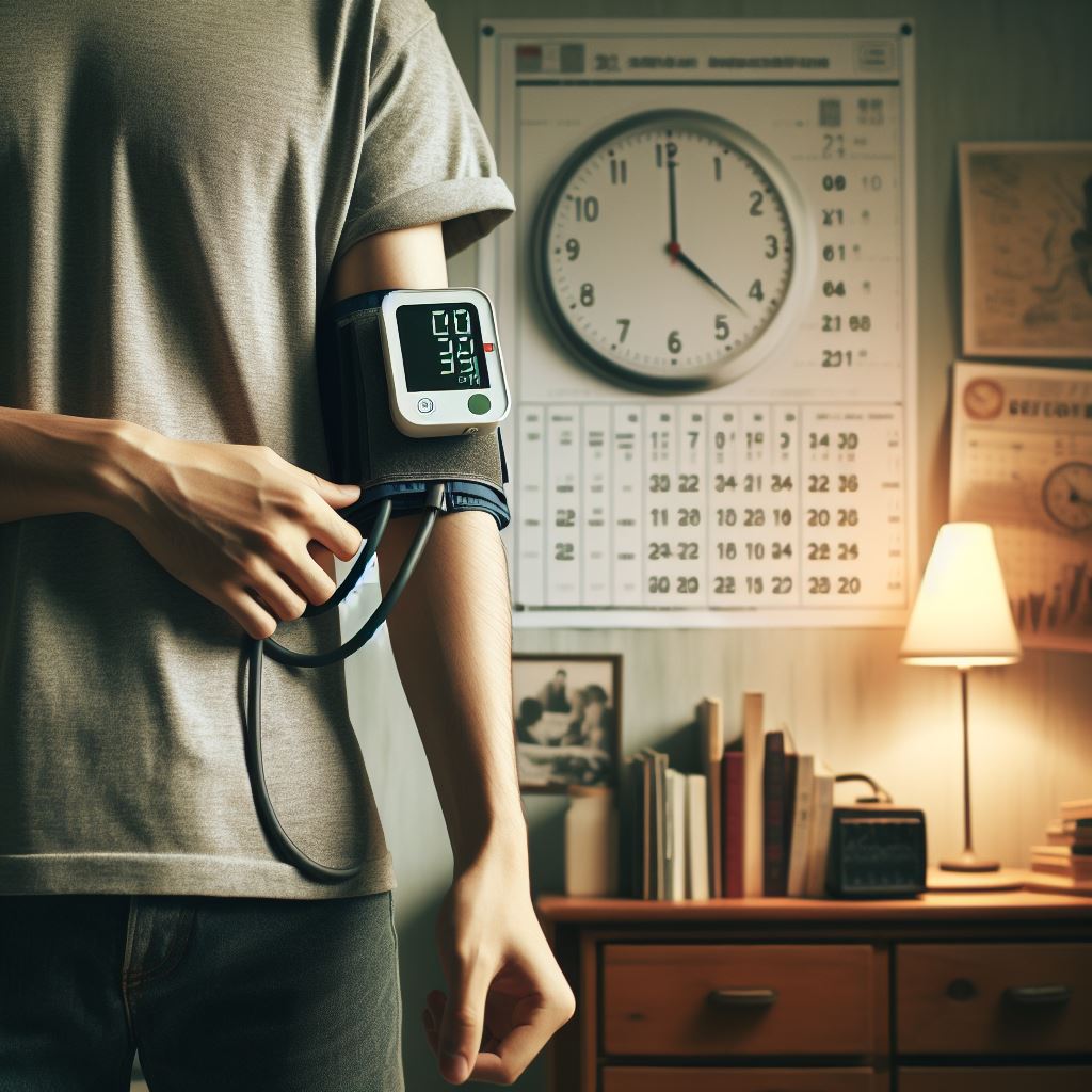 Ambulatory Blood Pressure Monitoring
-
 CT Imaging
-
 Sudden Cardiac Arrest